

By Dr. Pravin T. Goud
In fertility care, the journey of an egg does not always follow a straight or predictable path. On the day of egg retrieval, not every oocyte is developmentally ready. Some remain arrested in an immature state—seemingly silent, often overlooked, and frequently discarded. Yet many of these immature eggs are not inherently incapable. They simply require the right signals, environment, and time to reach their full potential.
My interest in in-vitro maturation (IVM) grew from a simple but persistent question: What does an immature human oocyte need in order to mature properly outside the body? And equally important—how closely can we recreate those conditions in the laboratory?
This research focused on two elements that repeatedly emerged as critical: the presence of cumulus cells and the role of epidermal growth factor (EGF). Together, they revealed how nuclear and cytoplasmic maturation are not isolated events, but interdependent processes shaped by communication, timing, and molecular support.
Why In-Vitro Maturation Matters
IVM offers an appealing alternative to conventional ovarian stimulation. It has the potential to reduce treatment costs, limit medication exposure, and eliminate the risk of ovarian hyperstimulation syndrome. For some patients—particularly those at medical risk or with polycystic ovarian features—IVM may represent a safer pathway to treatment.
Despite these advantages, pregnancy rates from in-vitro matured oocytes have historically lagged behind those from in-vivo matured eggs. This gap has often been attributed to incomplete or asynchronous maturation, particularly at the cytoplasmic level.
In other words, while an oocyte may reach metaphase II and appear mature, it may not yet possess the internal competence required for fertilization, chromosome segregation, or embryo development.
Understanding this distinction became central to our work.
Looking Beyond Nuclear Maturation
Meiosis is easy to observe. A polar body forms, the egg reaches metaphase II, and on the surface, maturation appears complete. But cytoplasmic maturation is more elusive. It involves redistribution of organelles, regulation of calcium signaling, cortical granule positioning, and molecular readiness for fertilization.
Many cleavage blocks observed in embryos derived from in-vitro matured oocytes likely arise not from chromosomal failure, but from subtle deficits in cytoplasmic preparedness.
This realization shaped our experimental design. We wanted to evaluate not only whether oocytes reached metaphase II, but whether they could activate, fertilize normally, and cleave after intracytoplasmic sperm injection (ICSI).
Why Cumulus Cells Deserve Attention
Cumulus cells are often treated as expendable—removed early in many laboratory protocols for convenience or visualization. Yet biologically, they serve as critical intermediaries between the oocyte and its environment.
These cells form gap junctions with the oocyte, enabling transfer of metabolites, signaling molecules, and regulatory factors. They also respond directly to gonadotrophins, relaying hormonal signals that the oocyte itself cannot fully perceive.
In vivo, the oocyte does not mature in isolation. Replicating that context in vitro is essential.
To understand the contribution of cumulus cells, we compared germinal vesicle (GV) oocytes matured with intact cumulus to those completely denuded prior to culture.
The Role of Epidermal Growth Factor
Epidermal growth factor is present in follicular fluid at physiologic concentrations, and its receptors are expressed on both oocytes and surrounding granulosa cells. In several animal models, EGF promotes meiotic progression and supports cytoplasmic maturation.
In humans, however, its role had not been clearly defined—particularly in relation to cumulus status and in hormonally supplemented, serum-free culture systems.
To address this, sibling GV oocytes were matured in defined media supplemented with gonadotrophins and estradiol, either with or without physiologic concentrations of EGF. This allowed direct comparison under tightly controlled conditions.
What We Observed During Maturation
The results were illuminating.
1. EGF Supports Nuclear Maturation in Denuded Oocytes
When cumulus cells were removed, EGF supplementation significantly increased the proportion of oocytes reaching metaphase II by 30 hours. This suggested that EGF can act directly on the oocyte, likely through its own receptors, to promote meiotic progression.
Without cumulus cells, the oocyte appears more dependent on direct molecular cues—and EGF filled that gap.
2. Cumulus Cells Improve Maturation Regardless of EGF
Oocytes matured with intact cumulus achieved higher rates of maturation overall, whether or not EGF was present. This confirms the intrinsic value of cumulus–oocyte communication during IVM.
Even in the absence of additional growth factors, cumulus cells enhanced synchrony and efficiency of nuclear maturation.
3. EGF Improves Cytoplasmic Competence in Cumulus-Intact Oocytes
The most telling results emerged after ICSI.
Among cumulus-intact oocytes, those matured with EGF demonstrated significantly higher rates of normal fertilization compared with those matured without it. This difference did not stem from nuclear maturation alone—it reflected improved cytoplasmic readiness.
In contrast, denuded oocytes showed similar activation rates but lower cleavage rates overall, highlighting the limits of nuclear maturation in isolation.
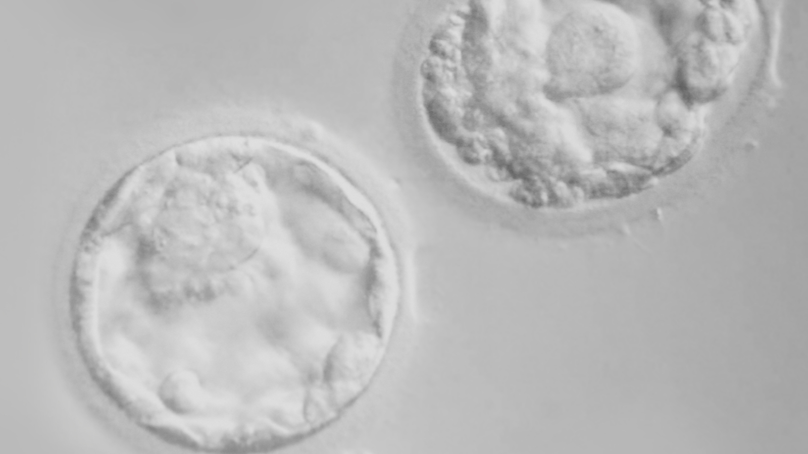
Cleavage as a Marker of Competence
Cleavage is a demanding biological test. It requires coordinated calcium signaling, intact cytoskeletal dynamics, and precise chromosome segregation. In our study, embryos derived from cumulus-intact oocytes cleaved at dramatically higher rates than those from denuded oocytes.
This finding reinforced a key message: oocytes mature better when they are not alone.
While EGF enhanced fertilization competence, the presence of cumulus cells was the dominant factor influencing early embryonic development.
Why Defined Culture Conditions Matter
One intentional aspect of this study was the exclusion of serum from the culture medium. Serum contains numerous undefined components, including growth factors, cytokines, and antioxidants that complicate interpretation.
By using albumin and clearly defined supplements, we were able to attribute observed effects specifically to EGF and cumulus status—rather than to unknown serum contaminants.
This approach also highlighted an important principle in reproductive science: understanding biology requires reducing noise, even when clinical practice sometimes relies on complexity.
Rethinking “Spare” Oocytes
A common misconception in IVF is that immature oocytes are inherently inferior. In reality, many GV-stage oocytes retrieved during stimulated cycles retain developmental potential—they simply lack optimal temporal cues.
Our findings suggest that with appropriate culture conditions, growth factor support, and preservation of cumulus cells, a meaningful proportion of these oocytes can mature, fertilize, and cleave.
This opens possibilities not only for research, but for thoughtful clinical application in selected contexts.
Clinical Implications Moving Forward
This work underscores several principles that continue to inform fertility care today:
- Nuclear maturation alone is insufficient to define egg quality
- Cumulus–oocyte communication remains critical in vitro
- Growth factors such as EGF modulate cytoplasmic readiness
- Culture systems matter—not just in composition, but in philosophy
Optimizing IVM is not about forcing eggs to mature faster, but about providing conditions that respect their biology.
Looking Ahead
In-vitro maturation remains a cutting-edge field—one that sits at the intersection of safety, accessibility, and biological precision. As we refine culture systems and deepen our understanding of oocyte–somatic cell interactions, the gap between in-vitro and in-vivo maturation continues to narrow.
The lesson this work taught me is simple but profound: maturation is not a switch. It is a conversation—between the oocyte, its supporting cells, and its environment. When we learn to listen carefully, outcomes improve.
About the Author
Dr. Pravin T. Goud is a reproductive endocrinologist, scientist, and clinician whose research focuses on oocyte quality and maturation, oxidative stress, gamete biology, and the molecular pathways governing fertilization and early embryo development. His published studies have contributed to a deeper scientific understanding of egg aging, cellular mechanisms influencing reproductive outcomes, and advances in in-vitro maturation systems and assisted reproductive technologies. Dr. Goud currently serves as Chief Scientific Officer at GenPrime, where he integrates scientific innovation with evidence-based fertility care and the clinical translation of reproductive biology research.